
If you have been suffering from blurred vision with halos and glares for years, then there is a possibility that you may have an ocular disease called keratoconus. But, what is keratoconus?
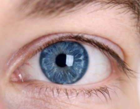
Keratoconus is an eye condition in which your cornea (the clear front surface of your eye that is shaped like a dome) thins and bulges outward into a cone shape. Your cornea is responsible for focusing light into your eye, but if it changes its shape, it brings light rays out of focus. This results in blurry and distorted vision. Eventually, you will find it hard to perform daily tasks such as driving and reading.
Are you curious and want to learn more about keratoconus? Here are the facts you will need to know.
What is keratoconus?
Our cornea is the clear, dome-shaped front surface at the front of our eye. It focuses light into our eyes. Keratoconus occurs when our normally dome-shaped (or round) cornea becomes thin and irregularly-shaped like a cone. The abnormal shape prevents light that enters the eye from being focused correctly on the retina, causing blurry and distorted vision.

There are two ways keratoconus can change your vision:
- When the cornea changes to a cone shape from its original rounded shape, the surface becomes distorted. This is known as irregular astigmatism.
- When the front of the cornea expands, vision becomes more nearsighted.
In the earliest stages, keratoconus only causes slight distortion and blurred vision, as well as increased sensitivity to glare and light. The first stages may appear in late teens or early 20s. The disease usually progresses for 10 to 20 years, and then the progress slows. It may affect each eye differently. As the disease progress, the cornea bulges more and your vision may become more distorted. Soft contact lenses or eyeglasses are usually used in order to correct mild astigmatism and nearsightedness caused by keratoconus in its early stages.
What causes keratoconus?
Research suggests that the weakening of the corneal tissue that results in keratoconus may be caused by an imbalance of enzymes within the cornea. This imbalance causing the cornea to become more susceptible to oxidative damage from compounds called free radicals. As a result, the cornea weakens and bulges forward.
It is not clear how this happens, but genetic and environmental factors are thought to be involved. 1 in about 10 people who have keratoconus also has a parent with the condition. Therefore, if you have it and have children, it is advisable that you check their eyes for keratoconus starting at age 10. The disease may also progress more rapidly in people with certain medical conditions, which include certain allergic conditions. The disease could also be related to chronic eye rubbing.
As mentioned before, keratoconus often starts in the teenage years and early 20s. however, it may also begin in childhood or in people up to age 30. It may occur in people at the age of 40 or older, but that is less common.
The changes in the shape of the cornea can happen rapidly or it may occur over the course of several years. The changes can stop at any time or continue for decades.
What are the symptoms of keratoconus?
The signs and symptoms of keratoconus may change as the disease progresses. The symptoms include:
- Distortion of objects in your vision
- Blurred or double vision
- Eye swelling
- Halos
- Eye redness or soreness
- Headaches
- Eyestrain
- Sudden worsening or clouding of vision
- Increased sensitivity to light and glare
- A need for frequent changes in eyeglass prescription
Can keratoconus complicate and damage vision?
In some situations, the cornea may swell rapidly, causing sudden reduced vision, as well as scarring of the cornea. A condition in which the inside lining of the cornea breaks down and caused rapid swelling as it allows fluid to enter the cornea.
Keratoconus may also advance. In this case, the cornea may become scarred, cause worsening vision problems that may need corneal transplant surgery.
While keratoconus does not typically result in complete blindness, it can decrease your vision to a level where you will find it difficult to perform normal daily activities.
How is keratoconus diagnosed?
Your doctor will review your medical and family history, as well as conducting an eye exam to diagnose keratoconus. Other tests to determine more details about the shape of your cornea may also be performed. Tests to diagnose keratoconus are as follows:
- Eye refraction – your eye doctor uses special equipment to evaluate your eyes to check for vision problems. You may be asked to look through a device containing wheels of different lenses, which will help judge the combination that gives you the sharpest vision. some doctors also use a hand-held instrument called a retinoscope to measure your eyes.
- Keratometry – during this test a circle of light is focused on your cornea and the reflection is measured to determine the shape of your cornea.
- Slit-lamp examination – this test involves directing a vertical light beam on the surface of your eye. Then, a low-powered microscope is used to view your eye. Your doctor then evaluates the shape of your cornea and checks for potential problems.
- Computerized corneal mapping – images of your cornea is recorded using special photographic tests, such as corneal topography and optical coherence tomography. The image is used to create a detailed shape map of the cornea’s surface. It can also measure the thickness of the cornea.
What are the treatment options for keratoconus?
The treatment for keratoconus varies depending on how quickly the condition is progressing and the severity of the condition. Mild to moderate forms of keratoconus can usually be treated with eyeglasses or soft contact lenses.
For some people with keratoconus, the cornea becomes scarred, making it difficult to wear contact lenses. If this happens, surgery may be needed.
Below are some treatment options for keratoconus:
Lenses
There are several types of lenses that can be used in the treatment for keratoconus, including:
- Eyeglasses or soft contact lenses – this is usually recommended for people with early stages of keratoconus. However, expect to change your prescription for eyeglasses or contact lens frequently as the shape of your cornea changes.
- Hard contact lenses – the next step in treating keratoconus that keeps on progressing is hard (rigid, gas permeable) contact lenses. These types of lenses can feel uncomfortable at first. However, many people are able to adjust wearing them and they provide excellent vision. Hard contact lenses can be made specifically to fit your cornea.
- Hybrid lenses – these types of lenses have a rigid center with a softer ring for increased comfort. Hybrid lenses are often used by those who cannot tolerate hard contact lenses.
- Piggyback lenses – rigid lenses can be uncomfortable. To make it more comfortable, your eye doctor may suggest “piggybacking” a hard contact lens on to of a soft one.
- Scleral lenses – this type of lenses can be used for very irregular shape changes in the cornea. Unlike traditional contact lenses that rest on the cornea, they sit on the white part of the eye and vault over the cornea without touching it.
Therapies
A type of therapy called corneal crosslinking can be a treatment option. This procedure strengthens your corneal tissue in order to halt bulging of the eye’s surface in keratoconus.
Corneal crosslinking has two versions:
- Epithelium-off crosslinking involves removing the outer layer of the cornea (known as the epithelium) to allow riboflavin (a type of vitamin B) to enter the cornea. The riboflavin is activated by UV light.
- Epithelium-on leaves the corneal epithelium intact during treatment.
Corneal cross-linking can help reduce the risk of progressive vision loss as it can stabilize the cornea at the early stages of the disease.
Surgery
If you experience corneal scarring, poor vision even with the strongest prescription lenses, extreme thinning of the cornea, or inability to wear contact lenses, surgery may be necessary. There are several types of surgery available, depending on the severity of the condition and the location of the bulge.
- Cornea transplant – if you experience extreme thinning of the cornea or corneal scarring, you may need a cornea transplant (also known as keratoplasty). There are several types of cornea transplants. The first one is called penetrating keratoplasty, which is a full cornea transplant. This procedure involves removing a full-thickness portion of the center of the cornea and replace it with donor tissue. The second one is called deep anterior lamellar keratoplasty (DALK), which preserves the inside lining of your cornea.
- Corneal inserts – this procedure involves placing tiny, clear, and crescent-shaped plastic inserts into the cornea in order to flatten the cone, improve vision, and support the cornea’s shape.